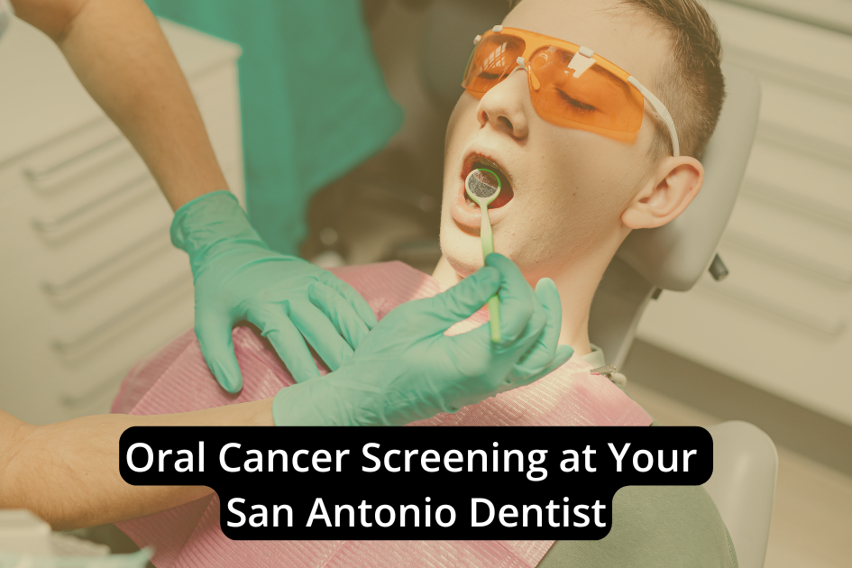
Think of an oral cancer screening as turning on a bright spotlight in a quiet room; we’re looking for what hides in plain sight. We’ll review your history, assess risk factors, inspect your mouth and throat under strong light, and palpate tissues for subtle firmness or asymmetry. We note sores, voice changes, or nonhealing areas with clinical precision. You’ll get clear next steps, sometimes on the spot, but knowing when further tests are warranted is the part most people don’t expect.
Why Early Detection Matters
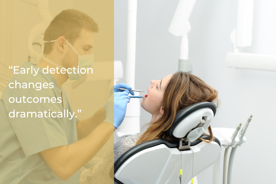
Although oral cancer can progress quietly, early detection changes outcomes dramatically. We prioritize detecting cellular and tissue changes before symptoms emerge because timing dictates prognosis. When we identify lesions early, survival exceeds 80%, and treatment stays less invasive. Our screening uses non‑invasive visualization and tactile assessment to map color, texture, and contour deviations that correlate with dysplasia or carcinoma in situ.
We deliver immediate feedback, explain findings, and, if needed, coordinate rapid referral for confirmatory evaluation. Evidence shows early-stage detection improves function preservation, reduces treatment morbidity, and lowers recurrence risk. With compassionate technique and advanced technology, we translate timely recognition into measurable, life‑saving advantages.
Who Should Consider Screening
With early detection established as the strongest predictor of outcomes, we recommend screening for all adults, with specific emphasis on risk tiers. Baseline: annual screening for everyone over 40. Elevated risk: tobacco or vaping use, heavy alcohol intake, prior oral lesions, HPV exposure, family history of head and neck cancer, or immunosuppression. Screen at least annually, often semiannually. Younger adults merit screening if they note persistent mouth sores, unexplained bleeding, color or texture changes, numbness, or chronic hoarseness. If you’ve had previous oral health concerns, maintain regular evaluations. Remember, absence of symptoms doesn’t exclude disease; routine screening drives survival gains.
What Happens at Your Initial Consultation
Before we begin the screening, we conduct a focused consultation to establish your risk profile and baseline. We review your medical and dental history, medications, tobacco and alcohol exposure, HPV status, prior lesions, and family history. We quantify risk, explain how it guides screening frequency, and outline next steps. You’ll know what we’re evaluating and why. We reserve the visual and tactile exam for the following phase, avoiding overlap here.
- Discuss personal and family cancer history
- Review lifestyle factors (tobacco, alcohol, sun, HPV)
- Catalog symptoms: sores, pain, numbness, voice changes
- Reconcile medications and systemic conditions
- Set screening interval and document baseline findings
Visual Examination of Oral Tissues
Having established your risk profile, we proceed to a systematic visual survey of the mouth and surrounding structures. We use bright, focused illumination and magnification to inspect lips, labial and buccal mucosa, gingiva, tongue surfaces, floor of mouth, hard and soft palate, and posterior oropharynx. We look for asymmetry, color changes (erythroplakia, leukoplakia), ulceration, nonhealing areas, abnormal vascular patterns, and borders that appear indurated or irregular. We may employ adjunctive light technologies to enhance contrast for subtle mucosal changes. You’ll breathe and swallow normally; the process is quick and noninvasive. Findings guide immediate feedback and, if indicated, evidence-based next steps.
Tactile Examination for Lumps and Irregularities
Next, we perform a methodical palpation of key structures to detect masses, induration, or tenderness that a visual exam can miss. We use gloved, moistened fingers to assess symmetry, texture, and mobility. You’ll feel gentle, deliberate pressure as we compare sides and note any fixed or firm areas that warrant attention. This step is quick, precise, and evidence-based.
- Floor of mouth: bidigital palpation to detect submucosal nodules
- Tongue: bimanual assessment for firmness or asymmetry
- Cheeks and lips: rolling technique for submucosal lesions
- Jaw and neck: lymph node mapping for enlargement or fixation
- Salivary glands: compression for tenderness, reduced flow, or firmness
Advanced Screening Technologies We Use
Building on our hands-on assessment, we apply adjunctive technologies to enhance detection sensitivity and specificity. We use fluorescence visualization to highlight abnormal mucosal changes that aren’t evident under white light. We supplement this with chemiluminescent rinses to accentuate surface irregularities and keratinization patterns. High-resolution intraoral imaging documents findings and enables precise comparisons over time. When indicated, we perform essential staining with toluidine blue to mark areas warranting closer evaluation. We integrate digital risk stratification using your history and exam data. These tools don’t replace clinical judgment; they refine it, helping us localize subtle changes earlier, with greater confidence and reproducibility.
Immediate Results and What They Mean
Once we complete the screening, we review your results with you immediately and classify findings as normal, low-suspicion, or requiring further evaluation. “Normal” means tissues look and feel healthy; no action beyond routine screening. “Low-suspicion” indicates minor irregularities we’ll monitor with targeted home care and a timed recheck. “Requires further evaluation” reflects distinct changes that merit prompt, focused assessment to clarify the cause.
- Normal: healthy color, texture, symmetry.
- Low-suspicion: mild color variation, transient irritation.
- Requires further evaluation: persistent ulcer, induration, fixation.
- Risk context: tobacco, alcohol, HPV history inform interpretation.
- Next steps: timeframe, self-check guidance, documentation provided.
When Further Evaluation Is Recommended
When a finding meets criteria for concern, we recommend further evaluation without delay. We act when lesions persist beyond two weeks, bleed easily, ulcerate, indurate, or display asymmetry, nonhealing white or red patches, unexplained pain, numbness, or a firm, fixed cervical node. High-risk history, tobacco, alcohol, and HPV lower our threshold.
Next steps are evidence-based. We photograph, document size, borders, color, and texture, then refer for adjunctive testing or biopsy, incisional or brush, through trusted local specialists. Imaging may assess nodes or bone. We coordinate timelines and communication, explain differential diagnoses, and emphasize that most abnormalities aren’t cancer, but timely pathology provides certainty.
Comfort, Safety, and Appointment Length

Comfort matters, and we design screenings to be painless, low-stress, and efficient. We use non-invasive techniques, visual and gentle tactile exams, to minimize discomfort while maximizing diagnostic value. Our protocols follow evidence-based guidelines and prioritize your safety with strict infection control and calibrated lighting and magnification. You’ll receive clear, immediate feedback, so you leave informed, not uncertain. Most visits take 60–90 minutes, including medical history review, examination, and discussion.
- No needles, no anesthesia
- Sterile instruments; single-use barriers
- Step-by-step explanation to reduce anxiety
- Thorough head, neck, and oral soft-tissue assessment
- Immediate summary with next-step recommendations (if needed)
How Often You Should Be Screened Based on Risk Factors
You’re informed about comfort and timing; now let’s calibrate screening frequency to your risk profile. We recommend annual screenings for most adults, especially those over 40. If you use tobacco, drink alcohol heavily, have HPV (especially HPV-16), a family history of head and neck cancer, prior dysplasia, or persistent oral lesions, we’ll increase frequency to every 3–6 months. Younger patients with high-risk factors follow the same intensified cadence. If you’ve had previous oral cancer, we coordinate three-month intervals initially, then extend as stable. New sores, color changes, numbness, or lumps warrant immediate evaluation. Early detection drives survival above 80–90%. We’ll individualize your schedule.
Conclusion
As we wrap up, remember that timely screening saves lives. Oral cancer can develop without pain or obvious symptoms in its early stages, which is why routine screenings are so important. During an oral cancer screening, we review risk factors, visually inspect and palpate oral tissues, document any abnormalities, and explain clear next steps when needed. If findings warrant further evaluation, we coordinate appropriate imaging or biopsy to ensure accurate diagnosis and timely care. Scheduling screenings at evidence-based intervals helps support early detection and better outcomes. Together, we can protect your oral health with precision, vigilance, and compassion.
Ready to take the next step? Book your oral cancer screening with Northwest Dental Center in San Antonio, TX, and let our team help you determine the right screening plan based on your individual risk and needs.